DICOM
Encord Computer Vision Glossary
DICOM, short for Digital Imaging and Communications in Medicine, is a standard format used in the medical industry for storing, transmitting, and sharing medical images and related data. It enables interoperability between different medical devices and systems, ensuring compatibility and consistency in image communication. DICOM files contain not only the image data but also metadata such as patient information, acquisition parameters, and image annotations. This standardization allows healthcare professionals to access and analyze medical images across different platforms and facilitates efficient collaboration and diagnosis. DICOM has revolutionized medical imaging, improving the efficiency and accuracy of healthcare delivery.
How is DICOM used in computer vision?
DICOM (Digital Imaging and Communications in Medicine) is primarily used in the medical field for storing and transmitting medical images. However, in the context of computer vision, DICOM plays a crucial role in enabling the integration of medical image data into computer vision pipelines and applications.
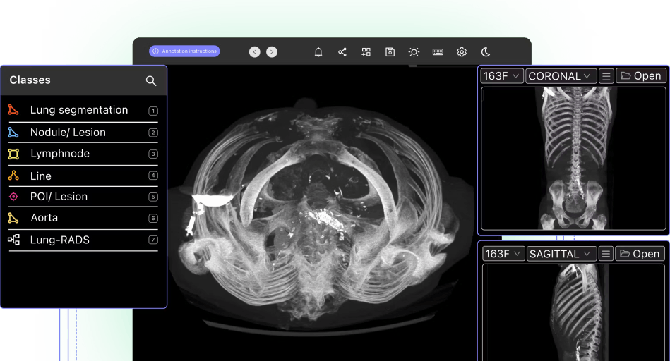
DICOM images, such as X-rays, CT scans, MRI scans, and ultrasound images, contain valuable visual information that can be analyzed and processed using computer vision techniques. By leveraging DICOM, computer vision algorithms can access and interpret the metadata associated with medical images, including patient demographics, acquisition parameters, and image annotations. This metadata provides critical context for understanding and analyzing the images effectively.
Computer vision algorithms applied to DICOM images can assist in various medical applications, such as image segmentation, object detection, image registration, disease diagnosis, and treatment planning. For example, computer vision models can be trained on labeled DICOM images to perform automated segmentation of organs or abnormalities, aiding in the identification and analysis of specific structures or regions of interest. Additionally, computer vision algorithms can help detect and classify anomalies, track disease progression, or assist in surgical planning based on DICOM image data.
What Advantages does DICOM offer in Computer Vision?
DICOM offers several advantages in computer vision. It provides a standardized format for storing and accessing medical image data, ensuring interoperability across different systems and devices. The metadata in DICOM images enhances the understanding and analysis of images. Additionally, DICOM integration facilitates collaboration between medical professionals and computer vision experts.
Can Computer Vision Models be Trained on DICOM images?
Yes, computer vision models can be trained on DICOM images. By leveraging labeled DICOM images, computer vision algorithms can learn to perform tasks like image segmentation, anomaly detection, and classification. Training on DICOM images allows models to learn from the rich visual information and associated metadata.