What’s the Difference Between DICOM and NIfTI?

Imaging standards and file formats play an essential role in medical imaging annotation. This article assesses the difference between DICOM and NIfTI, two of the most common medical imaging data formats.
One of the most significant advances in medical image annotation is the application of machine learning to evaluate images for a more precise, faster, and more accurate medical diagnosis.
Before machine learning (ML), artificial intelligence (AI), or any other diagnostic algorithms can be applied, you need to know that annotation software can handle the two most common medical and healthcare image file formats, including DICOM and NIfTI.
With medical images, the data type makes a huge difference. Unlike other image file formats (e.g., JPEG, PNG), healthcare professionals need to see A LOT more detail, so the raw data needs to be in a format that will reveal layers of the human body, organs, and the brain. Hence the need for 3D images or layers of 2D slices so that when it gets to the image processing stage, 3D visualizations, and rotations can be applied to get a much clearer understanding of the medical issue a doctor is trying to diagnose.
Computer vision and deep learning are playing an increasingly important role in the medical diagnosis and analysis process. Hence the advantage of specialist annotation and labeling tools that can handle DICOM data and NIfTI files so that segmentation and other labeling methods can be applied effectively.
Let’s review the difference between the DICOM and NIfTI, including who uses them and what they’re most commonly used for.
What is the DICOM standard?
The DICOM standard — Digital Imaging and Communications in Medicine (DICOM) — is used to exchange images and information and has been around for over over 20 years. The use of the DICOM format started to take off in the mid-nineties. DICOM comes with several support layers, allowing image senders and receivers to exchange information about the analyzed images.
Today, almost every imaging device used in radiology — including CT, MRI, Ultrasound, and RF — is equipped to support the DICOM standard. According to the standard facilitator, DICOM “enables the transfer of medical images in a multi-vendor environment and facilitates the development and expansion of picture archiving and communication systems.”
Imaging devices for other medical areas of expertise, including pathology, dermatology, endoscopy, neuroscience, and ophthalmology, are starting to use the DICOM standard.
Other layers of support include database connections and the ability for users to retrieve medical image header information, patient data, and metadata. Medical imaging devices can identify where images have been stored, making real-time retrieval and analysis easier. The third support layer includes image management, quality, storage, security, and patient scheduling information.
 One of the most important and valuable fundamentals of DICOM is the information model built into the standard. One of the most widely accepted definitions of this is:
“DICOM information objects are definitions of the information to be exchanged. One can think of them as templates that are reused over and over again when a modality generates a new image or other DICOM object. Each image type, and therefore information object, has specific characteristics.”
For example, “a CT image requires different descriptors in the image header than an ultrasound image or an ophthalmology image.”
Information objects are also known as Service Object Pairs (SOP) Classes. Each template, or SOP, comes with unique identifiers and a template so that when data is exchanged — whether that’s an annotated image or patient scheduling information — the two devices participating in the exchange transfer everything in as much detail as the respective users need.
Guidelines for the DICOM standard are overseen by the National Electrical Manufacturers Association (NEMA), known as the standard facilitator. Now let’s compare DICOM with another widely used medical and research imaging standard, NIfTI.
What is the NIfTI standard?
The Neuroimaging Informatics Technology Initiative (NIfTI) was established to work with medical and research device users and manufacturers to address some of the problems and shortfalls of other imaging standards. The NIfTI standard was specifically designed to address these challenges in the neuroimaging field, focusing on functional magnetic resonance imaging (fMRI).
One of the most significant challenges neurosurgeons faced with older image formats, specifically the Analyze 7.5 file format, was the lack of information about the orientation of image objects.
Orientation was ambiguous and unclear, forcing anyone attempting to analyze an image to add detailed notes about the orientation of objects within images. In particular, there was often confusion as to which side of the brain a doctor was looking at, a significant problem that needed a solution.
NIfTI solved this problem in the following way:“In the NIfTI format, the first three dimensions are reserved to define the three spatial dimensions — x, y and z —, while the fourth dimension is reserved to define the time points — t.” Other dimensions store “voxel-specific distributional parameters [and] vector-based data.”
According to NIfTI, “the primary goal of NIfTI is to provide coordinated and targeted service, training, and research to speed the development and enhance the utility of informatics tools related to neuroimaging.”
NIfTI has two standards, NIfTI-1 and NIfTI-2, with the second operating as a 64-bit improvement on the original. Not replacing it, but running alongside NIfTI-1, and supported by a wide range of neuroimaging medical devices and operating software systems. NIfTI is sponsored and overseen by The National Institute of Mental Health and the National Institute of Neurological Disorders and Stroke.
Now let’s review the differences between DICOM and NIfTI.
What are the differences between DICOM and NIfTI?
#1: Less metadata with NIfTI files
With a NIfTI file, you don’t need to populate the same number of tags (integer pairs) that DICOM image files required. There’s a lot less metadata to sift through and analyze; although in some respects, that’s a downside, as DICOM gives medical users layers of data about the image and patient.
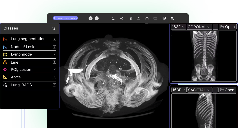
Metadata overlay being toggled in Encord
#2: DICOM files are often more cumbersome
DICOM is described as a “robust” standard, although that can cause difficulties for users. Strict formatting requirements govern DICOM transfers to ensure the receiving device supports SOP Classes and Transfer Syntaxes, e.g., the file format and encryption used to transfer data.
One device negotiates with another when DICOM files are being transferred. If one device can’t handle the information another device is attempting to send, “it will inform the requester so that the sender can either fall back to a different object (e.g., a previous version) or send the information to a different destination.”
Consequently, the handling, transfer, reading, and writing of NIfTI files are usually easier and quicker than DICOM image files. It’s like comparing a text file (NIfTI) with a Word file (DICOM). One has more detail, whereas the other is simpler, smaller, and easier to use. In many cases, the extra layers of data prove invaluable for image analysis across various medical fields.
#3: More information can be stored on DICOM files
As mentioned above, DICOM files allow medical professionals to store more information across multiple layers. You can create structured reports and even freeze an image so that other clinicians and healthcare data scientists can clearly see what an opinion/recommendation is based on.
So, although DICOM files are sometimes harder to handle, the information stored is more sophisticated and applicable across a wider range of medical use cases.
Read more: Encord’s guide to medical imaging experiments and best practices for machine learning and computer vision. #4: DICOM works with 2D layers, whereas NIfTI can display 3D detail
With NIfTI files, images and other data is stored in a 3D format. It’s specifically designed this way to overcome spatial orientation challenges of other medical image file formats.
On the other hand, DICOM image files and the associated data is made up of 2D layers. This allows you to view different slices of an image, which is especially useful when analyzing the human body and various organs.
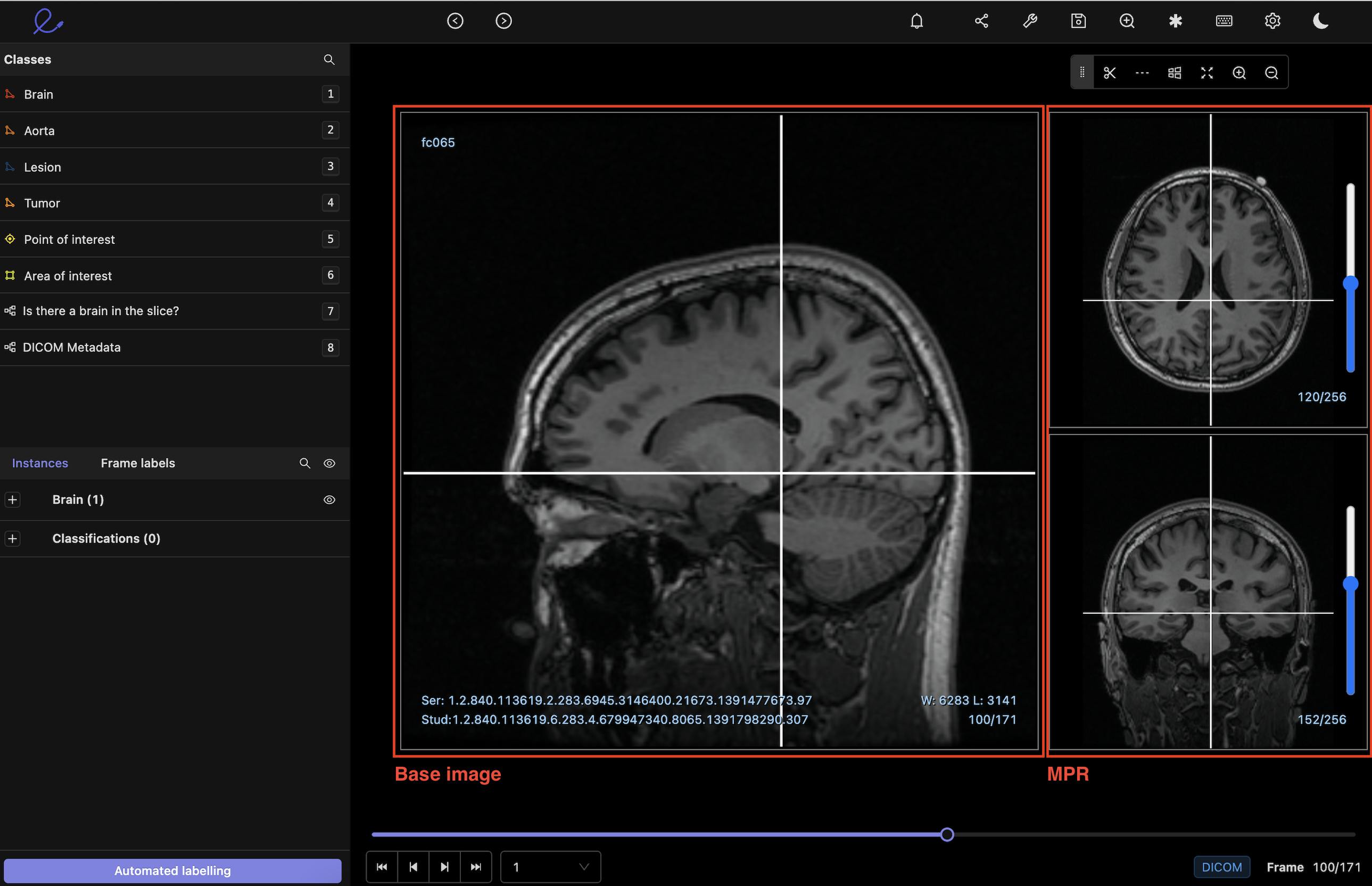
2D Multiplanar Reconstruction (MPR) in Encord
#5: NIfTI can take longer to load; DICOM enables users to display one layer at a time
Although the transfer of DICOM files often takes longer, NIfTI can take longer to load because the image data is stored in a 3D format. Depending on the software or image viewer being used, everything attempts to load at the same time. Whereas, once a DICOM file is received, users can display data one image at a time or simply load a single image and the associated metadata, such as the medial notes connected to that image.
The 2D layering of DICOM files means it’s quicker and easier to apply medical image annotation software or to send an image and notes to the printer in a medical workplace.
#6: You can convert DICOM into NIfTI
And finally — not a difference, but it’s useful to know healthcare professionals and data scientists can convert DICOM into NIfTI files without losing image quality. A NIfTI conversion is helpful when you need the data in a different format. Several open-source and proprietary software packages will help you do this, as the main challenge to overcome is combining the layers into a single image without losing any annotations, labels, or metadata.
Need to know more about annotating DICOM into NIfTI files? Check out: How to Annotate DICOM and NIfTI Files Who uses DICOM and what’s it used for?
DICOM is a more widely used image format in the healthcare profession, with a wide range of specialist fields deploying it, including radiology (for CT, X-Ray, MRI, Ultrasound, and RF), pathology, dermatology, endoscopy, and ophthalmology.
Hospitals worldwide use DICOM not only for the ability to share images and annotations but for the easy transfer and link to patient medical records, scheduling, and other valuable medical metadata objects.
DICOM is especially useful when trying to detect cancers. When DICOM files are labeled with DICOM-compatible medical image annotation tools, annotators can train models to spot cancers faster and more effectively, making drastic improvements in early detection.
Who uses NIfTI and what’s it used for?
NIfTI was originally created to solve a serious spatial orientation problem in neuroimaging, focusing on functional magnetic resonance imaging (fMRI). With NIfTI, neurosurgeons can quickly identify image objects, such as the right or left side of the brain, in 3D. It’s an invaluable asset when analyzing human brain images, a notoriously difficult organ to assess and annotate.
Radiologists are also using NIfTI file formats, as are image-based computer vision research teams and AI startups in the medical sector seeking FDA approval.
AI-based Medical Image Annotation for DICOM and NIfTI Labeling
Both medical image file formats are incredibly useful and powerful. Both have advantages and disadvantages. The good news is, whether you use DICOM or NIfTI, or both, Encord’s medical imaging annotation suite supports both file formats and standards. Our imaging software is the first purpose-built 3D annotation tool for healthcare AI
Encord has developed this software in close collaboration with medical professionals and healthcare data scientists, giving you a powerful automated image annotation suite with precise, 3D annotation built-in, fully auditable images, and unparalleled efficiency.
Accelerate your DICOM and annotation, labeling, and quality assurance with Encord Active: the open-source active learning toolkit for computer vision.
Want to stay updated? Follow us on Twitter and LinkedIn for more content on computer vision, training data, and active learning.
Here are some examples of healthcare and medical imaging projects involving Encord:
- Floy, an AI company that helps radiologists detect critical incidental findings in medical images, reduced CT & MRI Annotation time with AI-assisted labeling
- RapidAI reduced MRI and CT Annotation time by 70% using Encord for AI-assisted labeling.
- Stanford Medicine cut experiment duration time from 21 to 4 days while processing 3x the number of images in 1 platform rather than 3
Further Reading: Healthcare and Medical Imaging Annotation
Medical Image Segmentation: A Complete Guide
3 ECG Annotation Tools for Machine Learning
Frequently asked questions
Unlike general annotation platforms, Encord is specifically designed for the medical field, focusing on DICOM and other medical imaging formats. This specialization ensures that users benefit from tools and features tailored to meet the unique challenges of medical image annotation, such as accuracy and regulatory compliance.
Using DICOM format provides several advantages, including high-resolution rendering of signals and the ability to annotate waveform periods accurately. Encord allows for pixel spacing to be included in DICOM files, enabling automatic extraction of measurements like phase and frequency directly from the files.
Unlike traditional DICOM annotation tools that can be cumbersome and underutilized due to data import challenges, Encord is built specifically for ease of use in clinical settings. It allows for seamless integration with existing workflows and provides a more user-friendly experience for radiologists.
DICOM and STL exports are critical for users who require precise 3D models for surgical planning. Encord offers robust support for these formats, ensuring that users can seamlessly integrate and export their segmented models for further use in advanced implant surgeries.
Encord allows users to import and filter DICOM files using associated metadata, which can include date-time fields, text fields, number fields, and enum fields. This functionality helps users narrow down their search results to find the most relevant files quickly.
Yes, Encord enables users to customize the user interface according to their specific annotation workflows. This includes the ability to arrange images, videos, and documents side by side, allowing teams to configure the layout that best suits their needs.
Yes, Encord can support NIfTI files by allowing users to specify structured tags during the import process. This ensures that even without header data for unique study identifiers, the files can be organized correctly based on user-defined tags.
Absolutely. Encord is designed to support ongoing research projects involving DICOM data. Its flexible platform allows researchers to utilize the tool for multiple phases of their projects, ensuring continuity and efficiency in data analysis.
Yes, Encord features a native DICOM renderer, allowing for efficient management and annotation of DICOM scans. This capability is essential for teams working in the medical imaging space, ensuring that the data is processed correctly.
Encord utilizes a script to plot ECG signals using Python and matplotlib, converting high-resolution images from PNG to DICOM format. This allows for detailed visualization of individual features in the signal over time.