Medical Image Segmentation
Encord Computer Vision Glossary
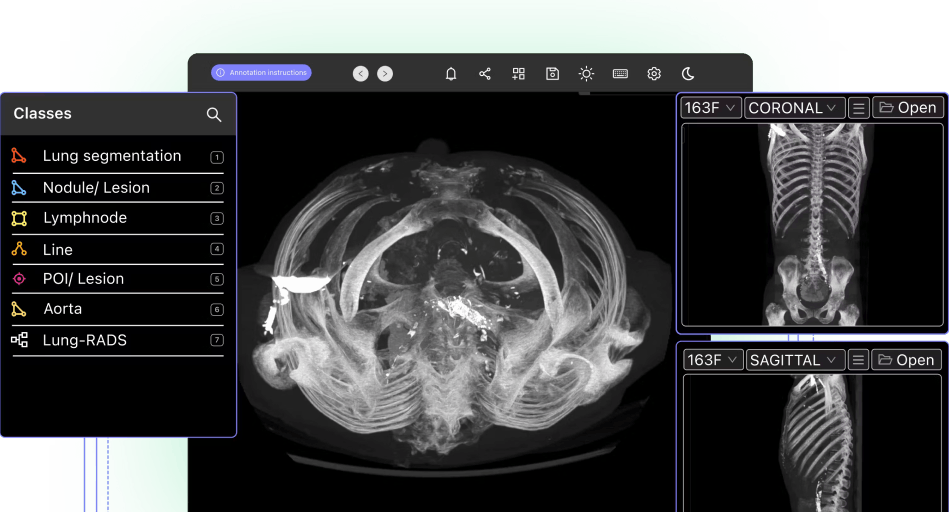
Medical image segmentation is a vital technique used in the field of healthcare to extract meaningful information from various imaging modalities such as magnetic resonance imaging (MRI), computed tomography (CT), and ultrasound. It plays a crucial role in clinical decision-making, treatment planning, and disease diagnosis. Here is an overview of medical image segmentation, its significance, and the methods used for accurate segmentation.
Importance of Medical Image Segmentation
Medical image segmentation is essential for precisely delineating anatomical structures and pathological regions of interest within medical images. It enables clinicians to isolate and analyze specific areas, facilitating accurate measurements, quantitative analysis, and treatment planning. Segmentation is particularly valuable in areas such as tumor delineation, organ volume estimation, tissue characterization, and surgical guidance. By segmenting medical images, clinicians gain crucial insights into patient conditions, leading to improved diagnosis, personalized treatment strategies, and better patient outcomes.

Methods Used in Medical Image Segmentation
Threshold-based Segmentation
This method involves setting a threshold value to distinguish different regions in an image based on intensity or color. It is a simple yet effective technique used for binary segmentation tasks, where the regions of interest can be separated from the background. However, it may not be suitable for complex structures or images with intensity variations.
Region-based Segmentation
This method involves partitioning an image into regions based on similarities in intensity, texture, or other features. Techniques such as region growing, watershed transform, and graph cut algorithms are commonly employed. Region-based segmentation is advantageous for images with uneven intensity distributions and can handle complex structures.
Edge-based Segmentation
Edge detection techniques, such as the Canny edge detector or Sobel operator, are used to identify boundaries between different regions. This method relies on detecting abrupt changes in pixel intensity and is useful for segmenting structures with clear boundaries. However, it may struggle with noisy or low-contrast images.
Machine Learning-based Segmentation
With the advent of deep learning, convolutional neural networks (CNNs) have revolutionized medical image segmentation. Models like U-Net, SegNet, and Mask R-CNN have shown remarkable performance in segmenting medical images. These models learn from large datasets and can handle complex structures, achieving high accuracy and robustness.

Challenges and Future Directions
Medical image segmentation still faces several challenges. Variability in image acquisition, noise, artifacts, and inter-patient anatomical variations can impact the accuracy of segmentation algorithms. Addressing these challenges requires further research in developing robust algorithms, incorporating multi-modal imaging, and exploring advanced techniques like 3D segmentation. Additionally, the integration of artificial intelligence and machine learning approaches holds promise for improving segmentation accuracy and efficiency.